产品名称
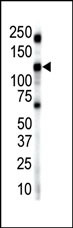
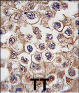
Insulin Receptor R Rabbit Polyclonal Antibody (N-term)
别名
Insulin receptor-related protein, IRR, IR-related receptor, Insulin receptor-related protein alpha chain, Insulin receptor-related protein beta chain, INSRR, IRR
存储缓冲液
Purified polyclonal antibody supplied in PBS with 0.09% (W/V) New?type?preservative?N. This antibody is prepared by Saturated Ammonium Sulfate (SAS) precipitation followed by dialysis against PBS.
Human Gene ID
NP_055030.1
Human Swissprot No.
P14616
特异性
This Insulin Receptor R antibody is generated from rabbits immunized with a KLH conjugated synthetic peptide between 27-57 amino acids from the N-terminal region of human Insulin Receptor R.
运输及保存条件
Maintain refrigerated at 2-8°C for up to 2 weeks. For long term storage store at -20°C in small aliquots to prevent freeze-thaw cycles.
背景介绍
Protein kinases are enzymes that transfer a phosphate group from a phosphate donor, generally the g phosphate of ATP, onto an acceptor amino acid in a substrate protein. By this basic mechanism, protein kinases mediate most of the signal transduction in eukaryotic cells, regulating cellular metabolism, transcription, cell cycle progression, cytoskeletal rearrangement and cell movement, apoptosis, and differentiation. With more than 500 gene products, the protein kinase family is one of the largest families of proteins in eukaryotes. The family has been classified in 8 major groups based on sequence comparison of their tyrosine (PTK) or serine/threonine (STK) kinase catalytic domains. The tyrosine kinase (TK) group is mainly involved in the regulation of cell-cell interactions such as differentiation, adhesion, motility and death. There are currently about 90 TK genes sequenced, 58 are of receptor protein TK (e.g. EGFR, EPH, FGFR, PDGFR, TRK, and VEGFR families), and 32 of cytosolic TK (e.g. ABL, FAK, JAK, and SRC families).
细胞定位
Membrane; Single-pass type I membrane protein.
功能
Receptor with tyrosine-protein kinase activity. Functions as a pH sensing receptor which is activated by increased extracellular pH. Activates an intracellular signaling pathway that involves IRS1 and AKT1/PKB.