产品名称
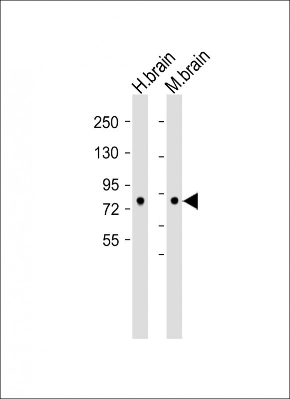
DCAMKL1 Rabbit Polyclonal Antibody (C-term)
别名
Serine/threonine-protein kinase DCLK1, Doublecortin domain-containing protein 3A, Doublecortin-like and CAM kinase-like 1, Doublecortin-like kinase 1, DCLK1, DCAMKL1, DCDC3A, KIAA0369
存储缓冲液
Purified polyclonal antibody supplied in PBS with 0.05% (V/V) Proclin 300. This antibody is purified through a protein A column, followed by peptide affinity purification.
Human Gene ID
NP_001182344.1;NP_001182345.1;NP_004725.1
Human Swissprot No.
O15075
特异性
This DCAMKL1 antibody is generated from rabbits immunized with a KLH conjugated synthetic peptide between 690-720 amino acids
of human DCAMKL1.
运输及保存条件
Maintain refrigerated at 2-8°C for up to 2 weeks. For long term storage store at -20°C in small aliquots to prevent freeze-thaw cycles.
背景介绍
Doublecortin-like kinase (DCAMKL1)(Ser/Thr protein kinase family) is essential for proper neurogenesis, neuronal migration, and axonal wiring. DCAMKL1 is involved in a calcium-signaling pathway controling neuronal migration in the developing brain, and participates in functions of the mature nervous system. DCAMKL1 protein shares high homology with doublecortin (DCX). DCLK, but not DCX, is highly expressed in regions of active neurogenesis in the neocortex and cerebellum. DCAMKL1 controls mitotic division by regulating spindle formation and also determines the fate of neural progenitors during cortical neurogenesis.
组织表达
In fetal tissues, highly expressed in brain, detectable in lung and liver, but not in kidney. In adult tissues, expressed ubiquitously in the brain, detectable in the heart, liver, spleen, thymus, prostate, testis, ovary, small intestine and colon. The type A isoforms seem to be expressed predominantly in fetal brain whereas type B isoforms are expressed abundantly in both fetal and adult brain.
功能
Probable kinase that may be involved in a calcium-signaling pathway controlling neuronal migration in the developing brain. May also participate in functions of the mature nervous system.