产品名称
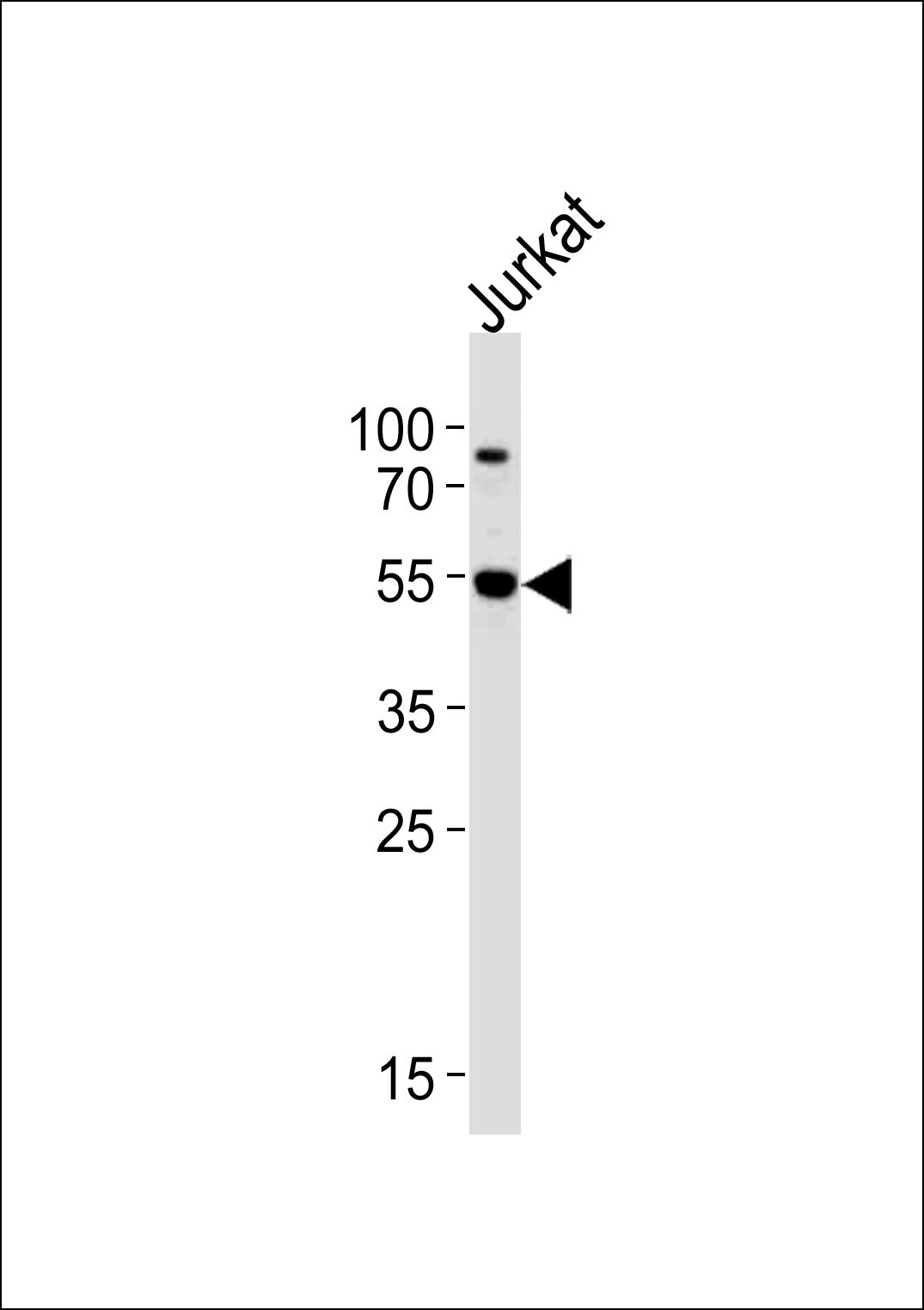
AKT3 Rabbit Polyclonal Antibody (Center)
别名
RAC-gamma serine/threonine-protein kinase, Protein kinase Akt-3, Protein kinase B gamma, PKB gamma, RAC-PK-gamma, STK-2, AKT3, PKBG
反应种属
Human, Rat, Monkey, Mouse
存储缓冲液
Purified polyclonal antibody supplied in PBS with 0.09% (W/V) New?type?preservative?N. This antibody is prepared by Saturated Ammonium Sulfate (SAS) precipitation followed by dialysis against PBS.
Human Gene ID
NP_001193658.1;NP_005456.1;NP_859029.1
Human Swissprot No.
Q9Y243
特异性
This AKT3 antibody is generated from rabbits immunized with a KLH conjugated synthetic peptide between 88-118 amino acids from the Central region of human AKT3.
运输及保存条件
Maintain refrigerated at 2-8°C for up to 2 weeks. For long term storage store at -20°C in small aliquots to prevent freeze-thaw cycles.
背景介绍
AKT3 is a member of the AKT, also called PKB, serine/threonine protein kinase family. AKT kinases are known to be regulators of cell signaling in response to insulin and growth factors. They are involved in a wide variety of biological processes including cell proliferation, differentiation, apoptosis, tumorigenesis, as well as glycogen synthesis and glucose uptake. This kinase has been shown to be stimulated by platelet-derived growth factor (PDGF), insulin, and insulin-like growth factor 1 (IGF1).
组织表达
In adult tissues, it is highly expressed in brain, lung and kidney, but weakly in heart, testis and liver. In fetal tissues, it is highly expressed in heart, liver and brain and not at all in kidney
细胞定位
Nucleus. Cytoplasm. Membrane; Peripheral membrane protein Note=Membrane-associated after cell stimulation leading to its translocation
功能
AKT3 is one of 3 closely related serine/threonine-protein kinases (AKT1, AKT2 and AKT3) called the AKT kinase, and which regulate many processes including metabolism, proliferation, cell survival, growth and angiogenesis. This is mediated through serine and/or threonine phosphorylation of a range of downstream substrates. Over 100 substrate candidates have been reported so far, but for most of them, no isoform specificity has been reported. AKT3 is the least studied AKT isoform. It plays an important role in brain development and is crucial for the viability of malignant glioma cells. AKT3 isoform may also be the key molecule in up-regulation and down-regulation of MMP13 via IL13. Required for the coordination of mitochondrial biogenesis with growth factor-induced increases in cellular energy demands. Down- regulation by RNA interference reduces the expression of the phosphorylated form of BAD, resulting in the induction of caspase- dependent apoptosis.