GTF2I Rabbit Polyclonal Antibody (C-term)
产品基本信息
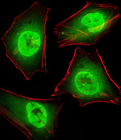
Fluorescent image of Hela cells stained with GTF2I Antibody (C-term). BD-PB4564 was diluted at 1:25 dilution. An Alexa Fluor 488-conjugated goat anti-rabbit lgG at 1:400 dilution was used as the secondary antibody (green). Cytoplasmic actin was counterstained with Alexa Fluor? 555 conjugated with Phalloidin (red).
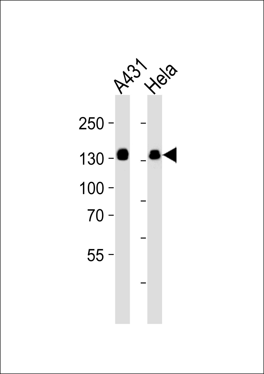
Western blot analysis of lysates from A431, Hela cell line (from left to right), using GTF2I Antibody (C-term)at 1:5000 dilution was used as the secondary antibody. Lysates at 35ug per lane.
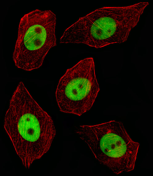
Fluorescent image of A549 cell stained with GTF2I Antibody (C-term)was used (1:400, 50 min at 37℃).Cytoplasmic actin was counterstained with Alexa Fluor? 555 (red) conjugated Phalloidin (7units/ml, 1 h at 37℃).GTF2I immunoreactivity is localized to Nucleus significantly.
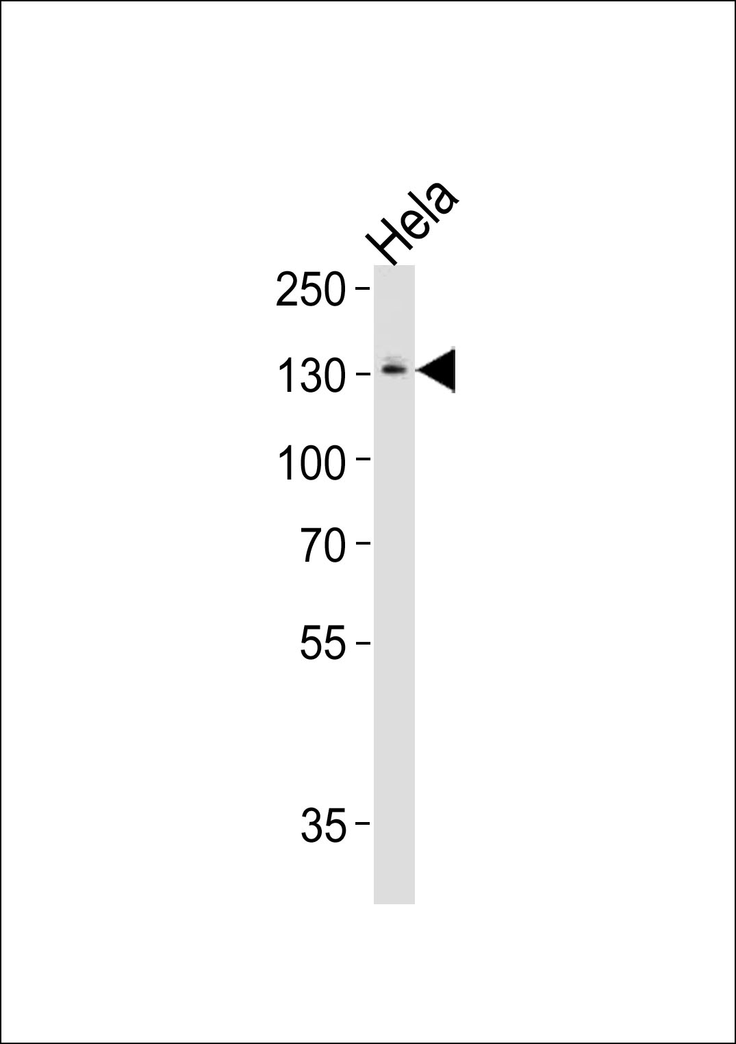
GTF2I Antibody (C-term) western blot analysis in Hela cell line lysates (35ug/lane).This demonstrates the GTF2I antibody detected the GTF2I protein (arrow).
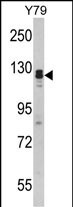
Western blot analysis of GTF2I Antibody (C-term) in Y79 cell line lysates (35ug/lane). GTF2I (arrow) was detected using the purified Pab.
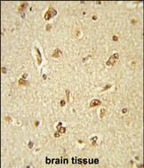
Formalin-fixed and paraffin-embedded human brain tissue reacted with GTF2I Antibody (C-term), which was peroxidase-conjugated to the secondary antibody, followed by DAB staining. This data demonstrates the use of this antibody for immunohistochemistry; clinical relevance has not been evaluated.
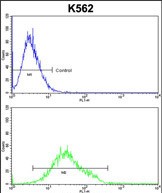
GTF2I Antibody (C-term) flow cytometric analysis of k562 cells (bottom histogram) compared to a negative control cell (top histogram).FITC-conjugated goat-anti-rabbit secondary antibodies were used for the analysis.
相关文献
产品问答
相关产品

市场:027-65023363 行政/人事:027-62439686 邮箱:marketing@brainvta.com 客服:18140661572(活动咨询、售后反馈等)
销售总监:张经理 18995532642 华东区:陈经理 18013970337 华南区:王经理 13100653525 华中/西区:杨经理 18186518905 华北区:张经理 18893721749
地址:中国武汉东湖高新区光谷七路128号中科开物产业园1号楼
Copyright © 武汉枢密脑科学技术有限公司. All RIGHTS RESERVED.
鄂ICP备2021009124号 DIGITAL BY VTHINK
