产品名称
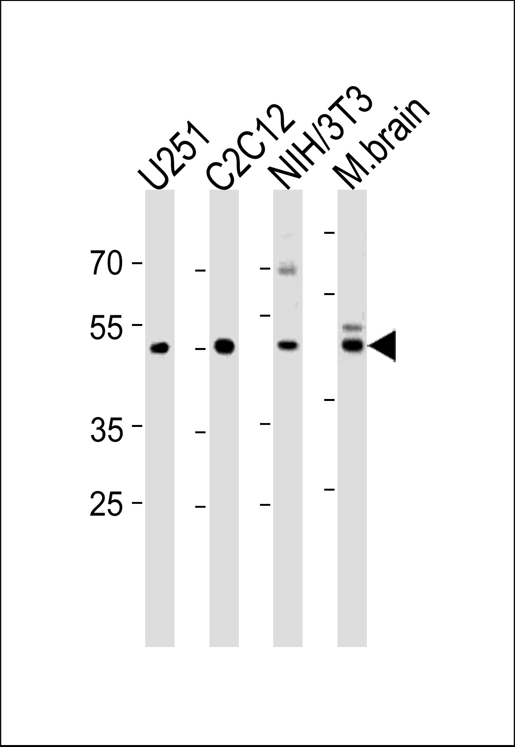
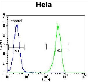
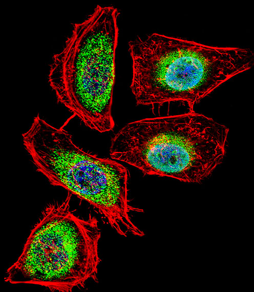
ENOA Rabbit Polyclonal Antibody (N-term)
别名
Alpha-enolase, 2-phospho-D-glycerate hydro-lyase, C-myc promoter-binding protein, Enolase 1, MBP-1, MPB-1, Non-neural enolase, NNE, Phosphopyruvate hydratase, Plasminogen-binding protein, ENO1, ENO1L1, MBPB1, MPB1
存储缓冲液
Purified polyclonal antibody supplied in PBS with 0.09% (W/V) New?type?preservative?N. This antibody is prepared by Saturated Ammonium Sulfate (SAS) precipitation followed by dialysis against PBS.
Human Gene ID
NP_001188412.1;NP_001419.1
Human Swissprot No.
P06733
特异性
This ENOA antibody is generated from rabbits immunized with a KLH conjugated synthetic peptide between 33-60 amino acids from the N-terminal region of human ENOA.
运输及保存条件
Maintain refrigerated at 2-8°C for up to 2 weeks. For long term storage store at -20°C in small aliquots to prevent freeze-thaw cycles.
背景介绍
ENO1 is one of three enolase isoenzymes found in mammals; the protein alpha-enolase, a homodimeric soluble enzyme, and is also a shorter monomeric structural lens protein, tau-crystallin. The two proteins are made from the same message. The full length protein, the isoenzyme, is found in the cytoplasm. The shorter protein is produced from an alternative translation start, is localized to the nucleus, and has been found to bind to an element in the c-myc promoter.
组织表达
The alpha/alpha homodimer is expressed in embryo and in most adult tissues. The alpha/beta heterodimer and the beta/beta homodimer are found in striated muscle, and the alpha/gamma heterodimer and the gamma/gamma homodimer in neurons
细胞定位
Cytoplasm. Cell membrane. Cytoplasm, myofibril, sarcomere, M line. Note=Can translocate to the plasma membrane in either the homodimeric (alpha/alpha) or heterodimeric (alpha/gamma) form. ENO1 is localized to the M line
功能
Glycolytic enzyme the catalyzes the conversion of 2- phosphoglycerate to phosphoenolpyruvate (PubMed:
29775581, PubMed:
1369209). In addition to glycolysis, involved in various processes such as growth control, hypoxia tolerance and allergic responses (PubMed:
2005901, PubMed:
10802057, PubMed:
12666133, PubMed:
29775581). May also function in the intravascular and pericellular fibrinolytic system due to its ability to serve as a receptor and activator of plasminogen on the cell surface of several cell-types such as leukocytes and neurons (PubMed:
12666133). Stimulates immunoglobulin production (PubMed:
1369209).