产品名称
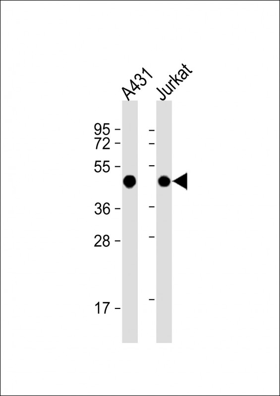
EBP1 Rabbit Polyclonal Antibody (Center)
别名
Proliferation-associated protein 2G4, Cell cycle protein p38-2G4 homolog, hG4-1, ErbB3-binding protein 1, PA2G4, EBP1
存储缓冲液
Purified polyclonal antibody supplied in PBS with 0.09% (W/V) New?type?preservative?N. This antibody is purified through a protein A column, followed by peptide affinity purification.
Human Gene ID
NP_006182.2
Human Swissprot No.
Q9UQ80
特异性
This EBP1 antibody is generated from rabbits immunized with a KLH conjugated synthetic peptide between 228-255 amino acids from the Central region of human EBP1.
运输及保存条件
Maintain refrigerated at 2-8°C for up to 2 weeks. For long term storage store at -20°C in small aliquots to prevent freeze-thaw cycles.
背景介绍
PA2G4 is an RNA-binding protein that is involved in growth regulation. This protein is present in pre-ribosomal ribonucleoprotein complexes and may be involved in ribosome assembly and the regulation of intermediate and late steps of rRNA processing. This protein can interact with the cytoplasmic domain of the ErbB3 receptor and may contribute to transducing growth regulatory signals. This protein is also a transcriptional co-repressor of androgen receptor-regulated genes and other cell cycle regulatory genes through its interactions with histone deacetylases. This protein has been implicated in growth inhibition and the induction of differentiation of human cancer cells.
组织表达
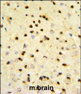
Isoform 2 is undetectable whereas isoform 1 is strongly expressed in cancer cells (at protein level). Isoform 1 and isoform 2 are widely expressed, including heart, brain, lung, pancreas, skeletal muscle, kidney, placenta and liver
细胞定位
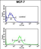
[Isoform 1]: Cytoplasm. Nucleus, nucleolus Note=Translocates to the nucleus upon treatment with HRG Phosphorylation at Ser-361 by PKC/PRKCD regulates its nucleolar localization.
功能
May play a role in a ERBB3-regulated signal transduction pathway. Seems be involved in growth regulation. Acts a corepressor of the androgen receptor (AR) and is regulated by the ERBB3 ligand neuregulin-1/heregulin (HRG). Inhibits transcription of some E2F1- regulated promoters, probably by recruiting histone acetylase (HAT) activity. Binds RNA. Associates with 28S, 18S and 5.8S mature rRNAs, several rRNA precursors and probably U3 small nucleolar RNA. May be involved in regulation of intermediate and late steps of rRNA processing. May be involved in ribosome assembly. Mediates cap- independent translation of specific viral IRESs (internal ribosomal entry site) (By similarity). Regulates cell proliferation, differentiation, and survival. Isoform 1 suppresses apoptosis whereas isoform 2 promotes cell differentiation (By similarity).