POLDIP3 Rabbit Polyclonal Antibody (N-term)
产品基本信息
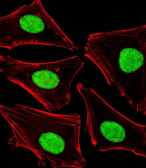
Fluorescent image of Hela cells stained with POLDIP3 Antibody (N-term). BD-PB4439 was diluted at 1:25 dilution. An Alexa Fluor 488-conjugated goat anti-rabbit lgG at 1:400 dilution was used as the secondary antibody (green). Cytoplasmic actin was counterstained with Alexa Fluor? 555 conjugated with Phalloidin (red).
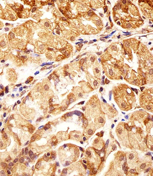
Immunohistochemical analysis of paraffin-embedded H. stomach section using POLDIP3 Antibody (N-term). BD-PB4439 was diluted at 1:25 dilution. A undiluted biotinylated goat polyvalent antibody was used as the secondary, followed by DAB staining.
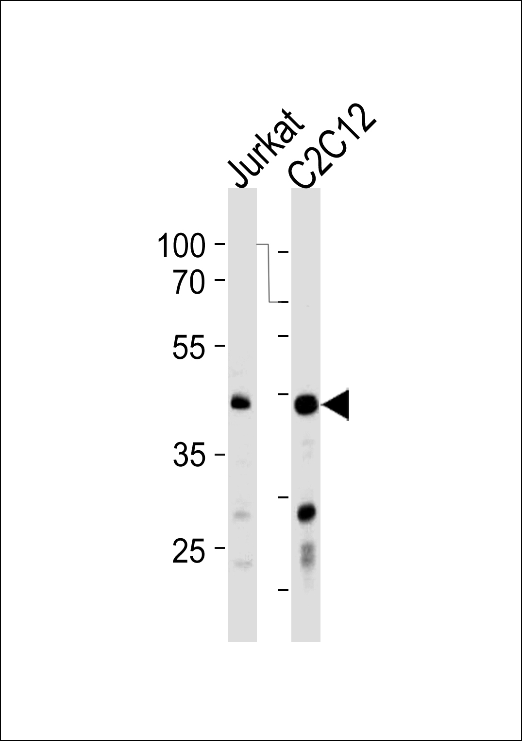
POLDIP3 Antibody (N-term) western blot analysis in Jurkat,mouse C2C12 cell line lysates (35ug/lane).This demonstrates the POLDIP3 Antibody antibody detected the POLDIP3 Antibody protein (arrow).
相关文献
产品问答
相关产品

市场:027-65023363 行政/人事:027-62439686 邮箱:marketing@brainvta.com 客服:18140661572(活动咨询、售后反馈等)
销售总监:张经理 18995532642 华东区:陈经理 18013970337 华南区:王经理 13100653525 华中/西区:杨经理 18186518905 华北区:张经理 18893721749
地址:中国武汉东湖高新区光谷七路128号中科开物产业园1号楼
Copyright © 武汉枢密脑科学技术有限公司. All RIGHTS RESERVED.
鄂ICP备2021009124号 DIGITAL BY VTHINK
