TARDBP Rabbit Polyclonal Antibody (N-term)
产品基本信息
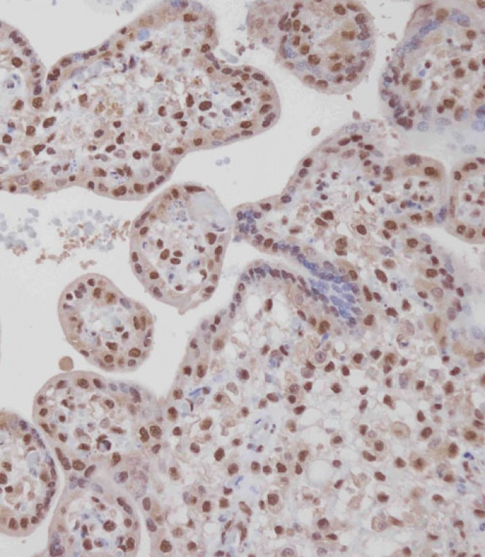
Immunohistochemical analysis of BD-PB4353 on paraffin-embedded Human placenta tissue. Tissue was fixed with formaldehyde at room temperature. Heat induced epitope retrieval was performed by EDTA buffer (pH9. 0). Samples were incubated with primary antibody(1:100) for 1 hour at room temperature. Undiluted CRF Anti-Polyvalent HRP Polymer antibody was used as the secondary antibody.
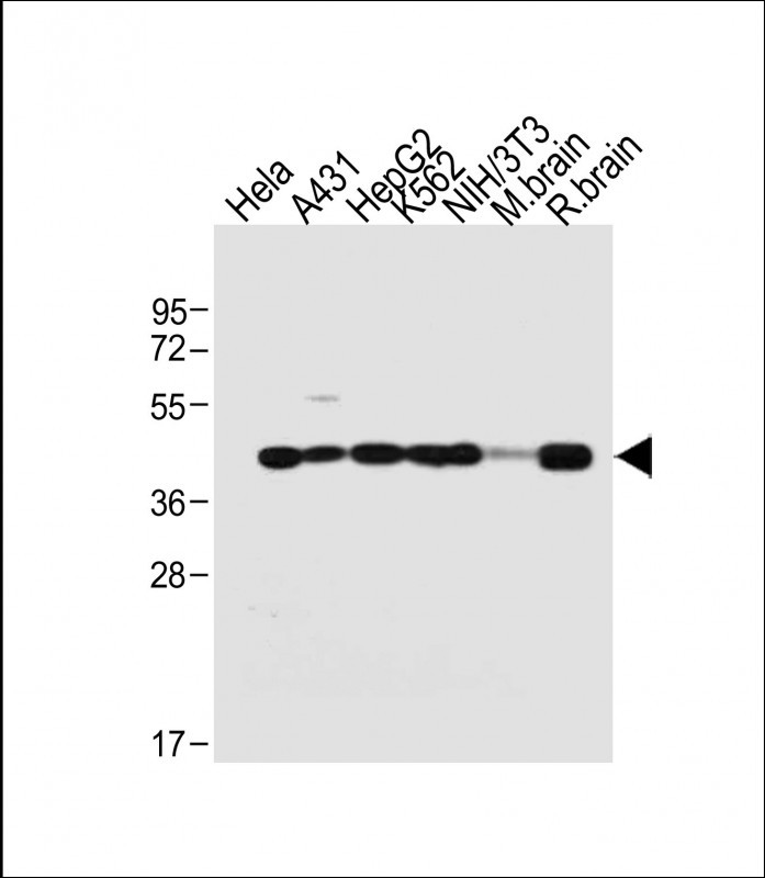
All lanes : Anti-TARDBP Antibody (N-term) at 1:2000 dilution Lane 1: Hela whole cell lysate Lane 2: A431 whole cell lysate Lane 3: HepG2 whole cell lysate Lane 4: K562 whole cell lysate Lane 5: NIH/3T3 whole cell lysate Lane 6: Mouse brain tissue lysate Lane 7: Rat brain tissue lysate Lysates/proteins at 20 μg per lane. Secondary Goat Anti-Rabbit IgG, (H+L), Peroxidase conjugated at 1/10000 dilution. Predicted band size : 45 kDa Blocking/Dilution buffer: 5% NFDM/TBST.
相关文献
产品问答
相关产品

市场:027-65023363 行政/人事:027-62439686 邮箱:marketing@brainvta.com 客服:18140661572(活动咨询、售后反馈等)
销售总监:张经理 18995532642 华东区:陈经理 18013970337 华南区:王经理 13100653525 华中/西区:杨经理 18186518905 华北区:张经理 18893721749
地址:中国武汉东湖高新区光谷七路128号中科开物产业园1号楼
Copyright © 武汉枢密脑科学技术有限公司. All RIGHTS RESERVED.
鄂ICP备2021009124号 DIGITAL BY VTHINK
