产品名称
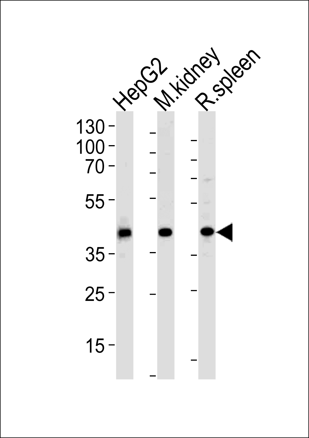
ACADL Rabbit Polyclonal Antibody (N-term)
别名
Long-chain specific acyl-CoA dehydrogenase, mitochondrial, LCAD, ACADL
存储缓冲液
Purified polyclonal antibody supplied in PBS with 0.09% (W/V) New?type?preservative?N. This antibody is purified through a protein A column, followed by peptide affinity purification.
Human Gene ID
NP_001599.1
Human Swissprot No.
P28330
特异性
This ACADL antibody is generated from rabbits immunized with a KLH conjugated synthetic peptide between 14-43 amino acids from the N-terminal region of human ACADL.
运输及保存条件
Maintain refrigerated at 2-8°C for up to 2 weeks. For long term storage store at -20°C in small aliquots to prevent freeze-thaw cycles.
背景介绍
The protein encoded by this gene belongs to the acyl-CoA
dehydrogenase family, which is a family of mitochondrial
flavoenzymes involved in fatty acid and branched chain amino-acid
metabolism. This protein is one of the four enzymes that catalyze
the initial step of mitochondrial beta-oxidation of straight-chain
fatty acid. Defects in this gene are the cause of long-chain
acyl-CoA dehydrogenase (LCAD) deficiency, leading to nonketotic
hypoglycemia.
细胞定位
Mitochondrion matrix {ECO:0000250|UniProtKB:P15650}
功能
Long-chain specific acyl-CoA dehydrogenase is one of the acyl-CoA dehydrogenases that catalyze the first step of mitochondrial fatty acid beta-oxidation, an aerobic process breaking down fatty acids into acetyl-CoA and allowing the production of energy from fats (By similarity). The first step of fatty acid beta-oxidation consists in the removal of one hydrogen from C-2 and C-3 of the straight-chain fatty acyl-CoA thioester, resulting in the formation of trans-2-enoyl- CoA (By similarity). Among the different mitochondrial acyl-CoA dehydrogenases, long-chain specific acyl-CoA dehydrogenase can act on saturated and unsaturated acyl-CoAs with 6 to 24 carbons with a preference for 8 to 18 carbons long primary chains (PubMed:
8823175, PubMed:
21237683).