TIMP2 (1O19) Mouse Monoclonal Antibody
产品基本信息
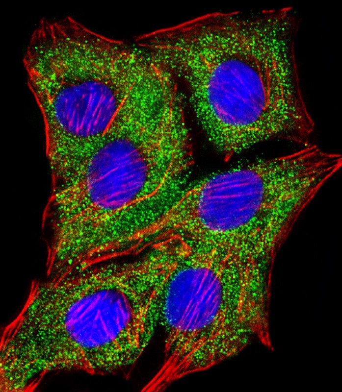
Immunofluorescent analysis of 4% paraformaldehyde-fixed, 0.1% Triton X-100 permeabilized A549 (human lung adenocarcinoma epithelial cell line) cells labeling TIMP2 with BD-PB4121 at 1/25 dilution, followed by Dylight? 488-conjugated goat anti-mouse IgG secondary antibody at 1/200 dilution (green). Immunofluorescence image showing cytoplasm staining on A549 cell line. Cytoplasmic actin is detected with Dylight? 554 Phalloidin at 1/100 dilution (red).The nuclear counter stain is DAPI (blue).

Immunohistochemical analysis of paraffin-embedded Human kidney section using Pink1. BD-PB4121 was diluted at 1:200 dilution. A undiluted biotinylated goat polyvalent antibody was used as the secondary, followed by DAB staining.
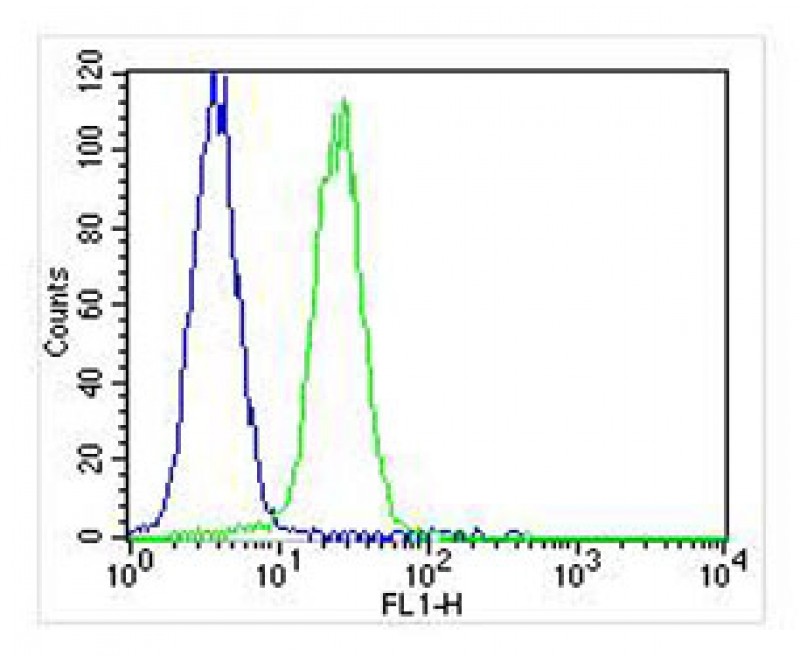
Overlay histogram showing K562 cells stained with BD-PB4121 (green line). The cells were fixed with 2% paraformaldehyde (10 min) and then permeabilized with 90% methanol for 10 min. The cells were then icubated in 2% bovine serum albumin to block non-specific protein-protein interactions followed by the antibody for 60 min at 37oC. The secondary antibody used was Goat-Anti-Mouse IgG, DyLight? 488 Conjugated Highly Cross-Adsorbed at 1/400 dilution for 40 min at 37oC. Isotype control antibody (blue line) was mouse IgG1 (1μg/1x10^6 cells) used under the same conditions. Acquisition of >10, 000 events was performed.
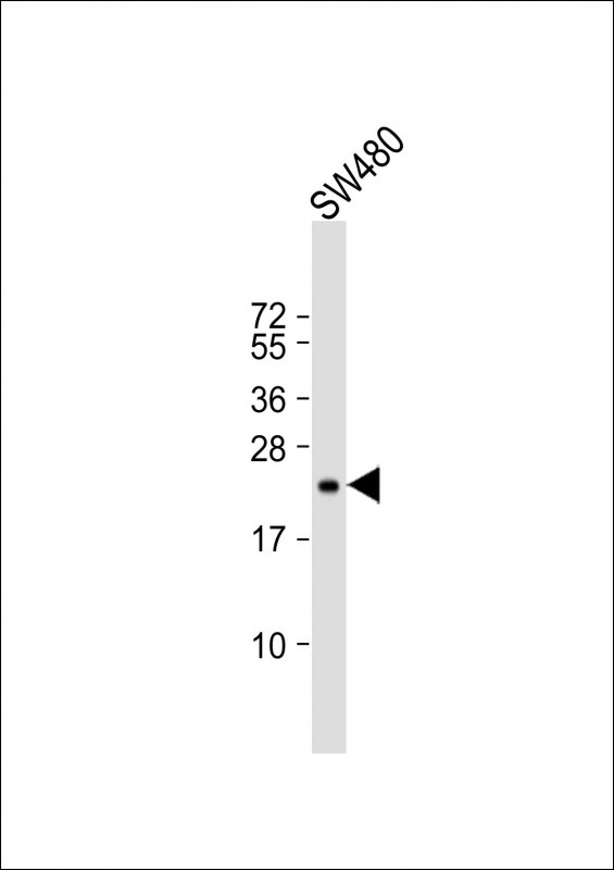
Anti-TIMP2 Antibody at 1:500 dilution + SW480 whole cell lysate Lysates/proteins at 20 μg per lane. Secondary Goat Anti-mouse IgG, (H+L), Peroxidase conjugated at 1/10000 dilution. Predicted band size : 24 kDa Blocking/Dilution buffer: 5% NFDM/TBST.
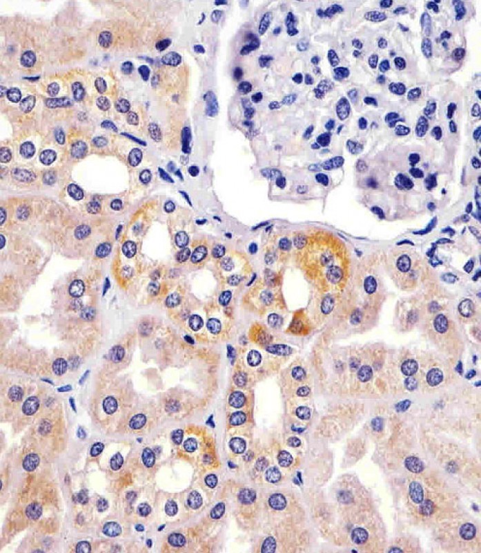
BD-PB4121 staining TIMP2 in human kidney sections by Immunohistochemistry (IHC-P - paraformaldehyde-fixed, paraffin-embedded sections). Tissue was fixed with formaldehyde and blocked with 3% BSA for 0. 5 hour at room temperature; antigen retrieval was by heat mediation with a citrate buffer (pH6). Samples were incubated with primary antibody (1/25) for 1 hours at 37°C. A undiluted biotinylated goat polyvalent antibody was used as the secondary antibody.
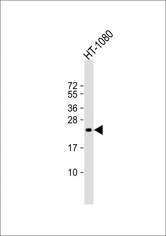
Anti-TIMP2 Antibody at 1:2000 dilution + HT-1080 whole cell lysate Lysates/proteins at 20 μg per lane. Secondary Goat Anti-mouse IgG, (H+L), Peroxidase conjugated at 1/10000 dilution. Predicted band size : 24 kDa Blocking/Dilution buffer: 5% NFDM/TBST.
相关文献
产品问答
相关产品

市场:027-65023363 行政/人事:027-62439686 邮箱:marketing@brainvta.com 客服:18140661572(活动咨询、售后反馈等)
销售总监:张经理 18995532642 华东区:陈经理 18013970337 华南区:王经理 13100653525 华中/西区:杨经理 18186518905 华北区:张经理 18893721749
地址:中国武汉东湖高新区光谷七路128号中科开物产业园1号楼
Copyright © 武汉枢密脑科学技术有限公司. All RIGHTS RESERVED.
鄂ICP备2021009124号 DIGITAL BY VTHINK
