Cn|En

现货抗体产品库

KIT (6C9) Mouse Monoclonal Antibody

产品详情 相关文献 产品问答 相关产品

产品基本信息

产品货号
BD-PB3883
产品名称
KIT (6C9) Mouse Monoclonal Antibody
别名
Mast/stem cell growth factor receptor Kit, SCFR, Piebald trait protein, PBT, Proto-oncogene c-Kit, Tyrosine-protein kinase Kit, p145 c-kit, v-kit Hardy-Zuckerman 4 feline sarcoma viral oncogene homolog, CD117, KIT, SCFR
类别
常规抗体
推荐应用
WB
反应种属
Human
Human Gene ID
NP_000213.1;NP_001087241.1
Human Swissprot No.
P10721
特异性
This KIT Antibody is generated from mouses immunized with human KIT recombinant protein.
稀释度
IHC-P-Leica~~1:1000;IF~~1:10~50;WB~~1:500~1:1000
预测分子量
110kDa
同种型
IgM
背景介绍
This gene encodes the human homolog of the proto-oncogene c-kit. C-kit was first identified as the cellular homolog of the feline sarcoma viral oncogene v-kit. This protein is a type 3 transmembrane receptor for MGF (mast cell growth factor, also known as stem cell factor). Mutations in this gene are associated with gastrointestinal stromal tumors, mast cell disease, acute myelogenous lukemia, and piebaldism. Multiple transcript variants encoding different isoforms have been found for this gene.
组织表达
[Isoform 3]: In testis, detected in spermatogonia in the basal layer and in interstitial Leydig cells but not in Sertoli cells or spermatocytes inside the seminiferous tubules (at protein level) (PubMed:20601678). Expression is maintained in ejaculated spermatozoa (at protein level) (PubMed:20601678)
细胞定位
[Isoform 1]: Cell membrane; Single-pass type I membrane protein [Isoform 3]: Cytoplasm. Note=Detected in the cytoplasm of spermatozoa, especially in the equatorial and subacrosomal region of the sperm head.
期货
现货

Immunohistochemical analysis of paraffin-embedded human appendix tissue using BD-PB3883 performed on the Leica® BOND RXm. Tissue was fixed with formaldehyde at room temperature; antigen retrieval was by heat mediation with a EDTA buffer (pH9. 0). Samples were incubated with primary antibody (1:1000) for 1 hours at room temperature. A undiluted biotinylated CRF Anti-Polyvalent HRP Polymer antibody was used as the secondary antibody.

Immunofluorescent analysis of U251 cells, using KIT Antibody conjugated Phalloidin (red).

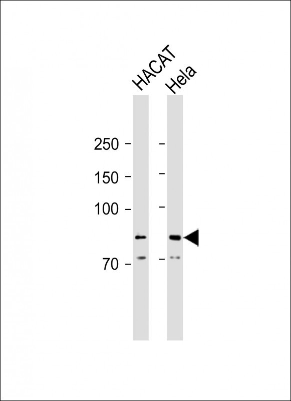
KIT Antibodywestern blot analysis in A549 cell line lysates (35μg/lane).This demonstrates the KIT antibody detected the KIT protein (arrow).

Fluorescent confocal image of an SY5Y cell stained with BD-PB3883 KIT antibody. SY5Y cells were fixed with 4% PFA (20 min), permeabilized with Triton X-100 (0.2%, 30 min), then incubated with BD-PB3883 KIT primary antibody (1:500, 2 h at room temperature). For secondary antibody, Alexa Fluor® 488 conjugated donkey anti-mouse antibody (green) was used (1:1000, 1h). Cytoplasmic actin was counterstained with Alexa Fluor® 555 (red) conjugated Phalloidin (5.25 μM, 25 min). Nuclei were counterstained with Hoechst 33342 (blue) (10 μg/ml, 3 min). Note the highly specific localization of the KIT immunosignal to the membranes, especially the plasma membrane. ibid. References for SY5Y : 1. Ross RA, et al. Coordinate morphological and biochemical interconversion of human neuroblastoma cells. J. Natl. Cancer Inst. 71: 741-749, 1983. [PubMed: 6137586]; 2. Biedler JL, et al. Multiple neurotransmitter synthesis by human neuroblastoma cell lines and clones. Cancer Res. 38: 3751-3757, 1978. [PubMed: 29704]. Ibid.

相关文献

产品问答

相关产品

免责声明| 法律支持| 联系方式

市场:027-65023363   行政/人事:027-62439686   邮箱:marketing@brainvta.com  

销售总监:张经理  18995532642  华东区:陈经理 18013970337   华南区:王经理 13100653525   华中/西区:杨经理 18186518905   华北区:张经理 18893721749

地址:中国武汉东湖高新区光谷七路128号中科开物产业园1号楼

Copyright © 武汉枢密脑科学技术有限公司. All RIGHTS RESERVED.
鄂ICP备2021009124号 DIGITAL BY VTHINK