TRAF2 (15L12) Mouse Monoclonal Antibody
产品基本信息
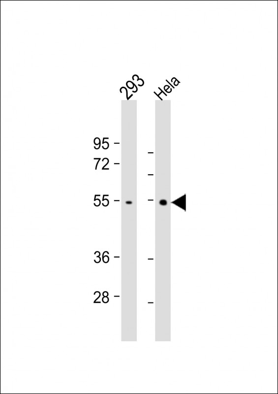
All lanes : Anti- at 1:1000 dilution Lane 1: 293 whole cell lysate Lane 2: Hela whole cell lysate Lysates/proteins at 20 μg per lane. Secondary Goat Anti-mouse IgG, (H+L), Peroxidase conjugated at 1/10000 dilution. Predicted band size : 56 kDa Blocking/Dilution buffer: 5% NFDM/TBST.
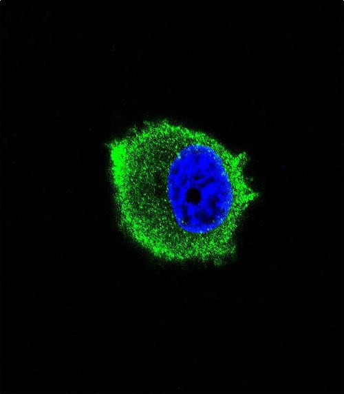
Confocal immunofluorescent analysis of TRAF2 Antibody with MCF-7 cell followed by Alexa Fluor? 488-conjugated goat anti-mouse lgG (green). DAPI was used to stain the cell nuclear (blue).
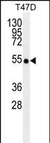
TRAF2/MB10188 antibody western blot analysis in T47D cell line lysates (35μg/lane).This demonstrates the TRAF2/MB10188 antibody detected the TRAF2/MB10188 protein (arrow).
相关文献
产品问答
相关产品

市场:027-65023363 行政/人事:027-62439686 邮箱:marketing@brainvta.com 客服:18140661572(活动咨询、售后反馈等)
销售总监:张经理 18995532642 华东区:陈经理 18013970337 华南区:王经理 13100653525 华中/西区:杨经理 18186518905 华北区:张经理 18893721749
地址:中国武汉东湖高新区光谷七路128号中科开物产业园1号楼
Copyright © 武汉枢密脑科学技术有限公司. All RIGHTS RESERVED.
鄂ICP备2021009124号 DIGITAL BY VTHINK
