产品名称
YES1(14A13)Mouse Monoclonal Antibody
别名
Tyrosine-protein kinase Yes, 2.7.10.2, Proto-oncogene c-Yes, p61-Yes, YES1, YES
Human Swissprot No.
P07947
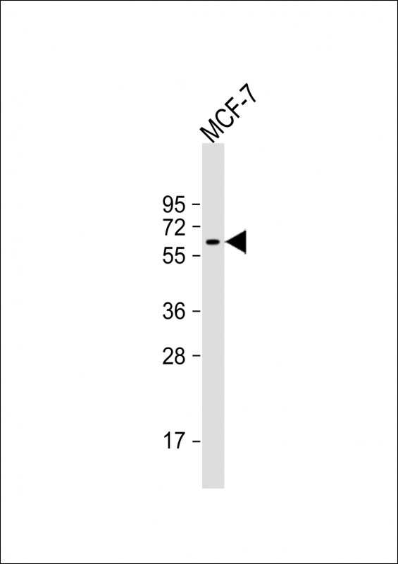
特异性
This YES1 antibody is generated from a mouse immunized with a recombinant protein of human YES1.
背景介绍
Non-receptor protein tyrosine kinase that is involved in the regulation of cell growth and survival, apoptosis, cell-cell adhesion, cytoskeleton remodeling, and differentiation. Stimulation by receptor tyrosine kinases (RTKs) including EGRF, PDGFR, CSF1R and FGFR leads to recruitment of YES1 to the phosphorylated receptor, and activation and phosphorylation of downstream substrates. Upon EGFR activation, promotes the phosphorylation of PARD3 to favor epithelial tight junction assembly. Participates in the phosphorylation of specific junctional components such as CTNND1 by stimulating the FYN and FER tyrosine kinases at cell-cell contacts. Upon T-cell stimulation by CXCL12, phosphorylates collapsin response mediator protein 2/DPYSL2 and induces T-cell migration. Participates in CD95L/FASLG signaling pathway and mediates AKT-mediated cell migration. Plays a role in cell cycle progression by phosphorylating the cyclin-dependent kinase 4/CDK4 thus regulating the G1 phase. Also involved in G2/M progression and cytokinesis.
组织表达
Expressed in the epithelial cells of renal proximal tubules and stomach as well as hematopoietic cells in the bone marrow and spleen in the fetal tissues. In adult, expressed in epithelial cells of the renal proximal tubules and present in keratinocytes in the basal epidermal layer of epidermis.
细胞定位
Cell membrane. Cytoplasm, cytoskeleton, microtubule organizing center, centrosome. Cytoplasm, cytosol Note=Newly synthesized protein initially accumulates in the Golgi region and traffics to the plasma membrane through the exocytic pathway