Cn|En

现货抗体产品库

SPDYA Rabbit Polyclonal Antibody (Center)

产品详情 相关文献 产品问答 相关产品

产品基本信息

产品货号
BD-PB2758
产品名称
SPDYA Rabbit Polyclonal Antibody (Center)
别名
Speedy protein A, Rapid inducer of G2/M progression in oocytes A, RINGO A, hSpy/Ringo A, Speedy-1, Spy1, SPDYA (HGNC:30613)
类别
常规抗体
推荐应用
WB
反应种属
Human
Human Gene ID
NP_001008779.1;NP_001136106.1;NP_877433.2
Human Swissprot No.
Q5MJ70
特异性
This SPDYA antibody is generated from rabbits immunized with a KLH conjugated synthetic peptide between 147-175 amino acids from the Central region of human SPDYA.
稀释度
WB~~1:1000;IHC-P~~1:100~500;FC~~1:10~50
预测分子量
36kDa
同种型
Rabbit Ig
背景介绍
SPDYA (speedy homolog A (Drosophila)) regulates the G1/S phase transition of the cell cycle by binding and activating CDC2, CDK2 and CDKN1B/KIP1. SPDYA can activate CDK2 without promoting CDK2 phosphorylation. SPDYA mediates cell survival during the DNA damage process through activation of CDK2.
组织表达
Highly expressed in testis. Expressed at a low level in wide range of tissues including bone marrow, brain, heart, kidney, colon, liver, placenta, spleen, skeletal muscle, salivary gland, thyroid gland, thymus, trachea and uterus. Expressed at a slightly higher level in adrenal gland, cerebellum, small intestine, lung, prostate and trachea. Expression is cell cycle-dependent, being restricted to cells in G1/S phase.
细胞定位
Nucleus
期货
现货
纯化
Purified polyclonal antibody supplied in PBS with 0.09% (W/V) sodium azide. This antibody is purified through a protein A column, followed by peptide affinity purification.

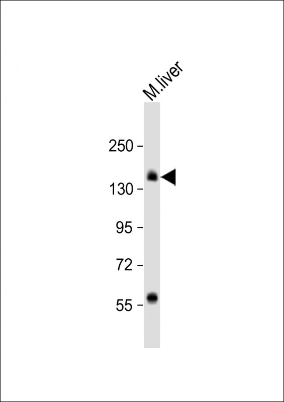
Anti-SPDYA Antibody (Center) at 1:1000 dilution + PC-3 whole cell lysate Lysates/proteins at 20 µg per lane. Secondary Goat Anti-Rabbit IgG, (H+L), Peroxidase conjugated at 1/10000 dilution. Predicted band size : 36 kDa Blocking/Dilution buffer: 5% NFDM/TBST.

SPDYA Antibody (Center) IHC analysis in formalin fixed and paraffin embedded prostate carcinoma followed by peroxidase conjugation of the secondary antibody and DAB staining. This data demonstrates the use of the SPDYA Antibody (Center) for immunohistochemistry. Clinical relevance has not been evaluated.

SPDYA Antibody (Center) flow cytometric analysis of K562 cells (right histogram) compared to a negative control cell (left histogram).FITC-conjugated goat-anti-rabbit secondary antibodies were used for the analysis.

Western blot analysis of SPDYA (arrow) using rabbit polyclonal SPDYA Antibody (Center). 293 cell lysates (2 ug/lane) either nontransfected (Lane 1) or transiently transfected (Lane 2) with the SPDYA gene.

相关文献

产品问答

相关产品

免责声明| 法律支持| 联系方式

市场:027-65023363   行政/人事:027-62439686   邮箱:marketing@brainvta.com  

销售总监:张经理  18995532642  华东区:陈经理 18013970337   华南区:王经理 13100653525   华中/西区:杨经理 18186518905   华北区:张经理 18893721749

地址:中国武汉东湖高新区光谷七路128号中科开物产业园1号楼

Copyright © 武汉枢密脑科学技术有限公司. All RIGHTS RESERVED.
鄂ICP备2021009124号 DIGITAL BY VTHINK