Cn|En

现货抗体产品库

GSTP1 Rabbit Polyclonal Antibody (Center)

产品详情 相关文献 产品问答 相关产品

产品基本信息

产品货号
BD-PB2661
产品名称
GSTP1 Rabbit Polyclonal Antibody (Center)
别名
Glutathione S-transferase P, GST class-pi, GSTP1-1, GSTP1, FAEES3, GST3
类别
常规抗体
推荐应用
WB
反应种属
Human, Rat, Mouse
Human Gene ID
NP_000843.1
Human Swissprot No.
P09211
特异性
This GSTP1 antibody is generated from rabbits immunized with a KLH conjugated synthetic peptide between 97-126 amino acids from the Central region of human GSTP1.
稀释度
WB~~1:1000;IF~~1:10~50;IHC~~1:2000;IHC-P~~1:100~500;FC~~1:10~50
预测分子量
23kDa
同种型
Rabbit Ig
背景介绍
Glutathione S-transferases (GSTs) are a family of enzymes that play an important role in detoxification by catalyzing the conjugation of many hydrophobic and electrophilic compounds with reduced glutathione. Based on their biochemical, immunologic, and structural properties, the soluble GSTs are categorized into 4 main classes: alpha, mu, pi, and theta. This GST family member is a polymorphic gene encoding active, functionally different GSTP1 variant proteins that are thought to function in xenobiotic metabolism and play a role in susceptibility to cancer, and other diseases.
细胞定位
Cytoplasm. Mitochondrion. Nucleus. Note=The 83 N-terminal amino acids function as un uncleaved transit peptide, and arginine residues within it are crucial for mitochondrial localization
期货
现货
纯化
Purified polyclonal antibody supplied in PBS with 0.09% (W/V) sodium azide. This antibody is purified through a protein A column, followed by peptide affinity purification.

GSTP1 Antibody (Center) western blot analysis in PC-3,Y79 cell line ,rat lung and testis tissue lysates (35ug/lane).This demonstrates the GSTP1 antibody detected the GSTP1 protein (arrow).

Fluorescent confocal image of A549 cell stained with GSTP1 Antibody (Center).A549 cells were fixed with 4% PFA (20 min), permeabilized with Triton X-100 (0.1%, 10 min), then incubated with GSTP1 primary antibody (1:25, 1 h at 37℃). For secondary antibody, Alexa Fluor® 488 conjugated donkey anti-rabbit antibody (green) was used (1:400, 50 min at 37℃).Cytoplasmic actin was counterstained with Alexa Fluor® 555 (red) conjugated Phalloidin (7units/ml, 1 h at 37℃). Nuclei were counterstained with DAPI (blue) (10 µg/ml, 10 min). GSTP1 immunoreactivity is localized to Cytoplasm and Mitochondria significantly.

Immunohistochemical analysis of paraffin-embedded Human tonsil section using Pink1. BD-PB2661 was diluted at 1:2000 dilution. A undiluted biotinylated goat polyvalent antibody was used as the secondary, followed by DAB staining.

Fluorescent confocal image of A549 cell stained with GSTP1 Antibody (Center).A549 cells were fixed with 4% PFA (20 min), permeabilized with Triton X-100 (0.1%, 10 min), then incubated with GSTP1 primary antibody (1:25, 1 h at 37℃). For secondary antibody, Alexa Fluor® 488 conjugated donkey anti-rabbit antibody (green) was used (1:400, 50 min at 37℃).Cytoplasmic actin was counterstained with Alexa Fluor® 555 (red) conjugated Phalloidin (7units/ml, 1 h at 37℃). Nuclei were counterstained with DAPI (blue) (10 µg/ml, 10 min). GSTP1 immunoreactivity is localized to Cytoplasm significantly.

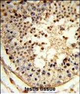
GSTP1 Antibody (Center) IHC analysis in formalin fixed and paraffin embedded human colon carcinoma followed by peroxidase conjugation of the secondary antibody and DAB staining. This data demonstrates the use of the GSTP1 Antibody (Center) for immunohistochemistry. Clinical relevance has not been evaluated.
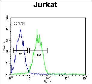
GSTP1 Antibody (Center) flow cytometric analysis of Ramos cells (right histogram) compared to a negative control cell (left histogram).FITC-conjugated goat-anti-rabbit secondary antibodies were used for the analysis.

相关文献

产品问答

相关产品

免责声明| 法律支持| 联系方式

市场:027-65023363   行政/人事:027-62439686   邮箱:marketing@brainvta.com  

销售总监:张经理  18995532642  华东区:陈经理 18013970337   华南区:王经理 13100653525   华中/西区:杨经理 18186518905   华北区:张经理 18893721749

地址:中国武汉东湖高新区光谷七路128号中科开物产业园1号楼

Copyright © 武汉枢密脑科学技术有限公司. All RIGHTS RESERVED.
鄂ICP备2021009124号 DIGITAL BY VTHINK