产品名称
SCYL1 Rabbit Polyclonal Antibody (N-term)
别名
N-terminal kinase-like protein, Coated vesicle-associated kinase of 90 kDa, SCY1-like protein 1, Telomerase regulation-associated protein, Telomerase transcriptional element-interacting factor, Teratoma-associated tyrosine kinase, SCYL1, CVAK90, GKLP, NTKL, TAPK, TEIF, TRAP
Human Gene ID
NP_001041683.1;NP_065731.3
Human Swissprot No.
Q96KG9
特异性
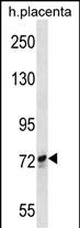
This SCYL1 antibody is generated from rabbits immunized with a KLH conjugated synthetic peptide between 156-185 amino acids from the N-terminal region of human SCYL1.
背景介绍
SCYL1 forms multimers following transfection into COS-7 cells. SCYL1 forms a 300-kD trimer using crosslinking reagents. Biochemical analysis revealed no phosphorylation or autophosphorylation activity.The 707-amino acid SCYL1 variant, variant 2, localized to centrosomes during mitosis. During interphase, fluorescence-tagged variant 2 localized in the cytoplasm as well as centrosomes. However, at the beginning of mitosis, the fluorescence appeared as a pair of bright nuclear foci that followed centrosome localization throughout mitosis, while maintaining diffuse cytoplasmic labeling. Endogenous variant 2 in HeLa cells showed a similar staining pattern. Centrosomal localization was independent of microtubules.
细胞定位
Cytoplasm, cytoskeleton, microtubule organizing center, centrosome. Endoplasmic reticulum-Golgi intermediate compartment Golgi apparatus, cis-Golgi network Note=Localized to the Endoplasmic reticulum-Golgi intermediate and cis- Golgi in an ARF1-independent manner [Isoform 2]: Cytoplasm. Note=Cytoplasmic throughout the cell cycle [Isoform 6]: Nucleus
纯化
Purified polyclonal antibody supplied in PBS with 0.09% (W/V) sodium azide. This antibody is purified through a protein A column, followed by peptide affinity purification.