Cn|En

现货抗体产品库

AGL Rabbit Polyclonal Antibody (C-term)

产品详情 相关文献 产品问答 相关产品

产品基本信息

产品货号
BD-PB2070
产品名称
AGL Rabbit Polyclonal Antibody (C-term)
别名
Glycogen debranching enzyme, Glycogen debrancher, 4-alpha-glucanotransferase, Oligo-1,4-1,4-glucantransferase, Amylo-alpha-1,6-glucosidase, Amylo-1,6-glucosidase, Dextrin 6-alpha-D-glucosidase, AGL, GDE
类别
常规抗体
推荐应用
WB
反应种属
Human
Human Gene ID
NP_000019.2;NP_000633.2;NP_000634.2;NP_000635.2;NP_000636.2;NP_000637.2
Human Swissprot No.
P35573
特异性
This AGL antibody is generated from rabbits immunized with a KLH conjugated synthetic peptide between 1479-1510 amino acids from the C-terminal region of human AGL.
稀释度
WB~~1:1000;IF~~1:10~50
预测分子量
175kDa
同种型
Rabbit Ig
背景介绍
AGL is a glycogen debrancher enzyme which is involved in glycogen degradation. This enzyme has two independent catalytic activities which occur at different sites on the protein: a 4-alpha-glucotransferase activity and a amylo-1,6-glucosidase activity. Mutations in the AGL gene are associated with glycogen storage disease although a wide range of enzymatic and clinical variability occurs which may be due to tissue-specific alternative splicing.
组织表达
Liver, kidney and lymphoblastoid cells express predominantly isoform 1; whereas muscle and heart express not only isoform 1, but also muscle-specific isoform mRNAs (isoforms 2, 3 and 4). Isoforms 5 and 6 are present in both liver and muscle
细胞定位
Cytoplasm. Note=Under glycogenolytic conditions localizes to the nucleus
期货
现货
纯化
Purified polyclonal antibody supplied in PBS with 0.09% (W/V) sodium azide. This antibody is purified through a protein A column, eluted with high and low pH buffers and neutralized immediately, followed by dialysis against PBS.

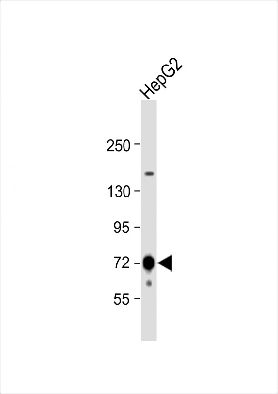
Anti-AGL Antibody (C-term) at 1:8000 dilution + human skeletal muscle lysate Lysates/proteins at 20 µg per lane. Secondary Goat Anti-Rabbit IgG, (H+L), Peroxidase conjugated at 1/10000 dilution. Predicted band size : 175 kDa Blocking/Dilution buffer: 5% NFDM/TBST.

Western blot using anti-AGL (C-term) antibody at 1:1000 dilution. A total of 20 ug of lysates was loaded for each tissue. Data courtesy of Dr. Alan Cheng, Department of Internal Medicine, Life Sciences Institute, University of Michigan Medical Center, Ann Arbor, Michigan.

Expression of myc-GS causes wild type but not the ÄCBD mutant of AGL to aggregate around the PAS-stain-positive inclusions. HepG2 cells were transfected with either HA-tagged wild-type AGL (HA-AGL) or HA-AGL ÄCBD. Cells were fixed in formalin and processed for IF using anti-HA (green) and anti-myc (red) antibodies. White arrows indicate colocalization of HA-AGL and myc-GS.

Confocal immunofluorescent analysis of AGL Antibody (C-term) with HepG2 cell followed by Alexa Fluor 488-conjugated goat anti-rabbit lgG (green). Actin filaments have been labeled with Alexa Fluor 555 phalloidin (red).DAPI was used to stain the cell nuclear (blue).

相关文献

产品问答

相关产品

免责声明| 法律支持| 联系方式

市场:027-65023363   行政/人事:027-62439686   邮箱:marketing@brainvta.com  

销售总监:张经理  18995532642  华东区:陈经理 18013970337   华南区:王经理 13100653525   华中/西区:杨经理 18186518905   华北区:张经理 18893721749

地址:中国武汉东湖高新区光谷七路128号中科开物产业园1号楼

Copyright © 武汉枢密脑科学技术有限公司. All RIGHTS RESERVED.
鄂ICP备2021009124号 DIGITAL BY VTHINK