产品名称
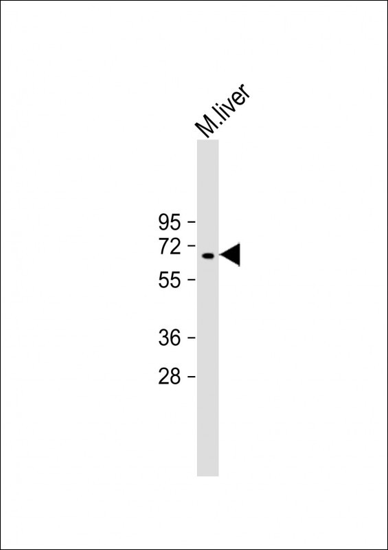
Mouse Camkk2 Rabbit Polyclonal Antibody (N-term)
别名
Calcium/calmodulin-dependent protein kinase kinase 2, CaM-KK 2, CaM-kinase kinase 2, CaMKK 2, Calcium/calmodulin-dependent protein kinase kinase beta, CaM-KK beta, CaM-kinase kinase beta, CaMKK beta, Camkk2, Kiaa0787
Human Swissprot No.
Q8C078
Mouse Gene ID
NP_001186605.1;NP_663333.1
特异性
This Mouse Camkk2 antibody is generated from rabbits immunized with a KLH conjugated synthetic peptide between 43-71 amino acids from the N-terminal region of mouse Camkk2.
背景介绍
Calcium/calmodulin-dependent protein kinase belonging to a proposed calcium-triggered signaling cascade involved in a number of cellular processes. Phosphorylates CAMK1, CAMK4 and CAMK1D (By similarity). Seems to be involved in hippocampal activation of CREB1.
组织表达
Expressed in all tissues tested. A differential expression pattern compared to CAMKK1 is observed in the brain
细胞定位
Nucleus {ECO:0000250|UniProtKB:Q96RR4}. Cytoplasm {ECO:0000250|UniProtKB:Q96RR4}. Cell projection, neuron projection {ECO:0000250|UniProtKB:Q96RR4}. Note=Predominantly nuclear in unstimulated cells, relocalizes into cytoplasm and neurites after forskolin induction. {ECO:0000250|UniProtKB:Q96RR4}
纯化
Purified polyclonal antibody supplied in PBS with 0.09% (W/V) sodium azide. This antibody is purified through a protein A column, followed by peptide affinity purification.