产品名称
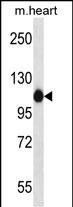
ACTN2 Rabbit Polyclonal Antibody (C-term)
别名
Alpha-actinin-2, Alpha-actinin skeletal muscle isoform 2, F-actin cross-linking protein, ACTN2
Human Gene ID
NP_001094.1
Human Swissprot No.
P35609
特异性
This ACTN2 antibody is generated from rabbits immunized with a KLH conjugated synthetic peptide between 751-779 amino acids from the C-terminal region of human ACTN2.
背景介绍
Alpha actinins belong to the spectrin gene superfamily
which represents a diverse group of cytoskeletal proteins,
including the alpha and beta spectrins and dystrophins. Alpha
actinin is an actin-binding protein with multiple roles in
different cell types. In nonmuscle cells, the cytoskeletal isoform
is found along microfilament bundles and adherens-type junctions,
where it is involved in binding actin to the membrane. In contrast,
skeletal, cardiac, and smooth muscle isoforms are localized to the
Z-disc and analogous dense bodies, where they help anchor the
myofibrillar actin filaments. This gene encodes a muscle-specific,
alpha actinin isoform that is expressed in both skeletal and
cardiac muscles.
组织表达
Expressed in both skeletal and cardiac muscle.
细胞定位
Cytoplasm, myofibril, sarcomere, Z line. Note=Colocalizes with MYOZ1 and FLNC at the Z-lines of skeletal muscle
纯化
Purified polyclonal antibody supplied in PBS with 0.09% (W/V) sodium azide. This antibody is purified through a protein A column, followed by peptide affinity purification.