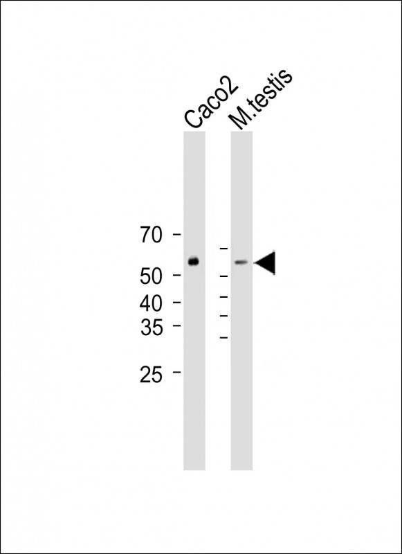
Cn|En

现货抗体产品库

AP1M1 Rabbit Polyclonal Antibody (Center)

产品详情 相关文献 产品问答 相关产品

产品基本信息

产品货号
BD-PB0913
产品名称
AP1M1 Rabbit Polyclonal Antibody (Center)
别名
AP-1 complex subunit mu-1, AP-mu chain family member mu1A, Adaptor protein complex AP-1 subunit mu-1, Adaptor-related protein complex 1 subunit mu-1, Clathrin assembly protein complex 1 mu-1 medium chain 1, Clathrin coat assembly protein AP47, Clathrin coat-associated protein AP47, Golgi adaptor HA1/AP1 adaptin mu-1 subunit, Mu-adaptin 1, Mu1A-adaptin, AP1M1, CLTNM
类别
常规抗体
推荐应用
WB
反应种属
Human, Rat, Mouse
Human Gene ID
NP_001123996.1;NP_115882.1
Human Swissprot No.
Q9BXS5
特异性
This AP1M1 antibody is generated from rabbits immunized with a KLH conjugated synthetic peptide between 205-234 amino acids from the Central region of human AP1M1.
稀释度
IF~~1:25;FC~~1:25;WB~~1:1000
预测分子量
49kDa
同种型
Rabbit Ig
背景介绍
The protein encoded by this gene is the medium chain of the trans-Golgi network clathrin-associated protein complex AP-1. The other components of this complex are beta-prime-adaptin, gamma-adaptin, and the small chain AP1S1. This complex is located at the Golgi vesicle and links clathrin to receptors in coated vesicles. These vesicles are involved in endocytosis and Golgi processing. Alternatively spliced transcript variants encoding distinct protein isoforms have been found for this gene. [provided by RefSeq].
细胞定位
Golgi apparatus. Cytoplasmic vesicle, clathrin- coated vesicle membrane; Peripheral membrane protein; Cytoplasmic side Note=Component of the coat surrounding the cytoplasmic face of coated vesicles located at the Golgi complex
期货
现货
纯化
Purified polyclonal antibody supplied in PBS with 0.09% (W/V) sodium azide. This antibody is purified through a protein A column, followed by peptide affinity purification.

Immunofluorescent analysis of 4% paraformaldehyde-fixed, 0. 1% Triton X-100 permeabilized U-251 MG cells labeling AP1M1 with BD-PB0913 at 1/25 dilution, followed by Dylight® 488-conjugated goat anti-Rabbit IgG secondary antibody at 1/200 dilution (green). Immunofluorescence image showing Cytoplasm and Weak Nucleus staining on U-251 MG cell line. Cytoplasmic actin is detected with Dylight® 554 Phalloidin(red). The nuclear counter stain is DAPI (blue).

Overlay histogram showing U-251 MG cells stained with BD-PB0913(green line). The cells were fixed with 2% paraformaldehyde (10 min) and then permeabilized with 90% methanol for 10 min. The cells were then icubated in 2% bovine serum albumin to block non-specific protein-protein interactions followed by the antibody for 60 min at 37ºC. The secondary antibody used was Goat-Anti-Rabbit IgG, DyLight® 488 Conjugated Highly Cross-Adsorbed at 1/200 dilution for 40 min at 37ºC. Isotype control antibody (blue line) was rabbit IgG1 (1μg/1x10^6 cells) used under the same conditions. Acquisition of >10, 000 events was performed.

All lanes : Anti-AP1M1 Antibody (Center) at 1:1000 dilution Lane 1: Human brain lysate Lane 2: PC-3 whole cell lysate Lane 3: U-251 MG whole cell lysate Lysates/proteins at 20 µg per lane. Secondary Goat Anti-Rabbit IgG, (H+L), Peroxidase conjugated at 1/10000 dilution. Predicted band size : 49 kDa Blocking/Dilution buffer: 5% NFDM/TBST.

相关文献

产品问答

相关产品

免责声明| 法律支持| 联系方式

市场:027-65023363   行政/人事:027-62439686   邮箱:marketing@brainvta.com  

销售总监:张经理  18995532642  华东区:陈经理 18013970337   华南区:王经理 13100653525   华中/西区:杨经理 18186518905   华北区:张经理 18893721749

地址:中国武汉东湖高新区光谷七路128号中科开物产业园1号楼

Copyright © 武汉枢密脑科学技术有限公司. All RIGHTS RESERVED.
鄂ICP备2021009124号 DIGITAL BY VTHINK