PSMA5(12D2)Mouse Monoclonal Antibody
产品基本信息
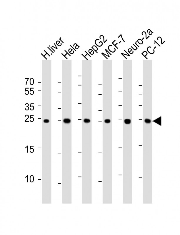
All lanes : Anti-PSMA5 Antibody at 1:1000 dilution Lane 1: human liver lysate Lane 2: Hela whole cell lysate Lane 3: HepG2 whole cell lysate Lane 4: MCF-7 whole cell lysate Lane 5: Neuro-2a whole cell lysate Lane 6: PC-12 whole cell lysate Lysates/proteins at 20 μg per lane. Secondary Goat Anti-mouse IgG, (H+L), Peroxidase conjugated at 1/10000 dilution. Predicted band size : 26 kDa. Blocking/Dilution buffer: 5% NFDM/TBST.
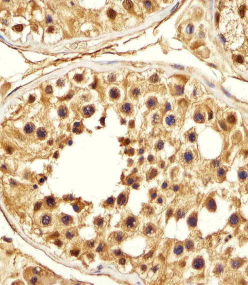
Immunohistochemical analysis of paraffin-embedded H. testis section using PSMA5 Antibody. BD-PB0712 was diluted at 1:25 dilution. A peroxidase-conjugated goat anti-mouse IgG at 1:400 dilution was used as the secondary antibody, followed by DAB staining.
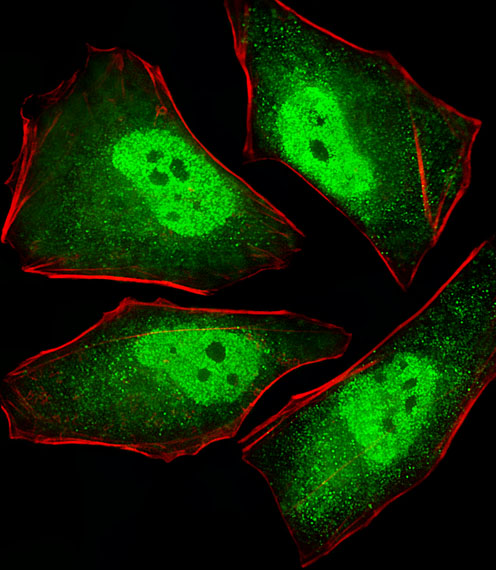
Fluorescent image of Hela cells stained with XAF1 PSMA5 Antibody. BD-PB0712 was diluted at 1:25 dilution. An Alexa Fluor? 488-conjugated goat anti-mouse lgG at 1:400 dilution was used as the secondary antibody (green). Cytoplasmic actin was counterstained with Alexa Fluor? 555 conjugated with Phalloidin (red).
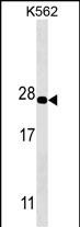
PSMA5 Antibody western blot analysis in K562 cell line lysates (35μg/lane).This demonstrates the PSMA5 antibody detected the PSMA5 protein (arrow).
相关文献
产品问答
相关产品

市场:027-65023363 行政/人事:027-62439686 邮箱:marketing@brainvta.com 客服:18140661572(活动咨询、售后反馈等)
销售总监:张经理 18995532642 华东区:陈经理 18013970337 华南区:王经理 13100653525 华中/西区:杨经理 18186518905 华北区:张经理 18893721749
地址:中国武汉东湖高新区光谷七路128号中科开物产业园1号楼
Copyright © 武汉枢密脑科学技术有限公司. All RIGHTS RESERVED.
鄂ICP备2021009124号 DIGITAL BY VTHINK
